Many natural adhesives and natural phenomena in nature provide valuable references for the development of adhesives/adhesive materials. Based on this, biomimetic adhesive materials, by mimicking the adhesion mechanisms of natural adhesives and utilizing biocompatible materials for their preparation, possess excellent biocompatibility and high adhesion properties. These materials overcome the limitations of existing tissue adhesives, including non-degradability, cytotoxicity, incompatibility with wet surfaces, and inability to adapt to dynamic tissue movements. These biomimetic adhesive materials can not only be used as wound dressings but also as medical glues to promote wound healing, serving as alternatives to conventional tissue sutures or tissue adhesives.
For instance, mussels in the ocean anchor themselves to various surfaces through adhesive proteins, enabling them to withstand the immense shear forces of ocean waves without being swept away. Slugs secrete mucus when confronted by predators, adhering themselves to rocks and soil surfaces to evade capture. Geckos, with their adhesive toe pads, can defy gravity and climb. These natural phenomena have inspired the development of biomimetic adhesive materials that harness similar mechanisms.
1. Mussels
Mussels, a type of marine organism widely distributed along coastal regions, adhere securely to various solid substrates in seawater through the mussel foot proteins (Mfp) secreted by their byssus glands. To date, scientists have identified six types of mussel foot proteins (Mfp1-Mfp6), all of which contain varying proportions of 3,4-dihydroxyphenylalanine (DOPA). Further research has revealed that the higher the DOPA content, the stronger the adhesive properties of Mfp. The key factor contributing to DOPA's adhesive properties is its catechol functional group, which not only readily forms hydrogen bonds with proteins but also exhibits robust metal coordination and chelating abilities.
Under alkaline, aerobic, or enzymatic conditions, the phenolic hydroxyl groups of DOPA can be oxidized into quinones or semiquinones. The oxidized catechol groups engage in Michael addition and Schiff base reactions with amino and thiol groups on proteins, undergo intramolecular cyclization to form dehydroindole structures, and undergo self-disproportionation reactions to generate radicals, which crosslink with substrates through coupling reactions. Additionally, catechol participates in other physicochemical interactions, such as hydrogen bonding, metal coordination, π-π or π-cation interactions, and hydrophobic interactions, further enhancing adhesion and cohesion with substrates.
Numerous researchers have conducted molecular simulations to develop a series of mussel-inspired medical adhesives. Some scholars have used catechol-modified ε-poly-L-lysine and oxidized dextran as the main chains, crosslinked in situ through Schiff base and catechol-Fe3? coordination to form a hydrogel bioadhesive. The unique dual-dynamic bond crosslinking structure endows the bioadhesive with higher mechanical strength and adhesion strength while maintaining rapid dissociation and excellent self-healing capabilities. Inspired by the mussel adhesion mechanism, other scholars have introduced protocatechualdehyde (PA), which contains catechol and aldehyde groups, and Fe3? into an adhesive composed of sodium alginate and gelatin. The interaction between alginic acid and the amino and carboxyl groups of gelatin imparts cohesion to the adhesive, which is further enhanced by the introduction of PA and Fe3?, accelerating the absorption and adhesion to substrates. This hydrogel exhibits sufficient mechanical strength and adhesiveness, effectively sealing wounds and promoting healing in in vivo skin incision experiments.
Furthermore, scientists have also developed artificial melanin-like biopolymers known as polydopamine (PDA). The reactive catechol groups and primary amine groups on PDA exhibit excellent adhesion properties, allowing it to adhere to almost all types of substrate surfaces. The catechol groups can also form covalent bonds with amino-terminal or thiol-terminal reagents through various chemical reactions, facilitating the attachment of small molecules, biomolecules, and polymers to the PDA surface. Additionally, PDA boasts biocompatibility, photothermal effects, antibacterial properties, drug-loading capabilities, and other advantages.
2.Barnacles
Like mussels, barnacles adhere to substrates through the secretion of adhesive proteins. The adhesive proteins secreted by barnacles are classified into primary cement and secondary cement. Primary cement is the glue-like substance secreted during normal growth and development of barnacle individuals, while secondary cement is re-secreted when external forces cause the barnacle's baseplate to detach from its attachment substrate. Although the two types of cement produced by barnacles share similar peptide segments, they differ in their degrees of self-assembly and curing cross-linking. Notably, catechol structures are absent in barnacle proteins, with only a small number of disulfide bonds present, indicating significant differences in the adhesion mechanisms between barnacles and mussels.
At present, the exact adhesion mechanism of barnacle cement remains unclear. Some researchers believe that chemical reactions play a role in the curing process, while others suggest that it is related to the assembly structure of the proteins. The barnacle adhesive protein cp19k, located at the interface, is considered crucial for adhesion to various surfaces, such as ship hulls, rocks, and shells of other marine organisms.
Using atomic force microscopy (AFM) imaging and force spectroscopy, researchers have studied the effect of pH on the self-assembly and adhesion properties of barnacle adhesive protein cp19k. They confirmed for the first time that the bacterially recombinant barnacle adhesive protein cp19k does not contain 3,4-dihydroxyphenylalanine (DOPA) or any other post-translationally modified amino acids. Furthermore, this recombinant protein can self-assemble into aggregated nanofibers under acidic pH conditions. Under moderately acidic conditions, the unassembled cp19k protein exhibits adhesion strength on mica that is only slightly lower than that of commercially available mussel adhesive protein mixtures, but its adhesion capability decreases significantly as pH increases. In contrast, pre-assembled nanofibers of cp19k maintain higher stability and show stronger adhesion in acidic and low-salinity conditions compared to those in alkaline and high-salinity seawater.
Moreover, other barnacle adhesive proteins also play indispensable roles in the adhesion process. For instance, the barnacle adhesive protein 52 kDa exhibits starch-like aggregation and self-assembly units, suggesting that the crosslinking points in barnacle cement are not covalent bonds but insoluble and resilient crosslinking networks generated by starch-like self-assembled protein aggregates. This starch-like self-assembly enhances the cohesion of the cement, contributing to its curing process.
3.Octopus
The octopus can adhere to surfaces in the ocean through its specialized sucker structures. Its adhesion mechanism differs from that of mussels and barnacles, as it does not rely on chemical reactions for adhesion but solely on the unique structure of its suckers. When the octopus comes into contact with an object, the muscles within its suckers attach to the surface, forming a seal at the outer edge. This creates a negative pressure within the cavity of the sucker, utilizing the pressure difference to achieve adhesion with the object.
When the internal and external pressures within the sucker cavity are equalized, the contraction of both transverse and longitudinal muscles triggers the release of circular muscle contractions. The contraction of transverse muscles causes the volume of the sucker's cavity to expand, resulting in a decrease in pressure within the cavity. This, in turn, enhances the suction and adhesion effect. Therefore, the octopus's adhesive force is primarily generated by the pressure difference created through the contraction of transverse muscles, allowing for efficient adhesion.
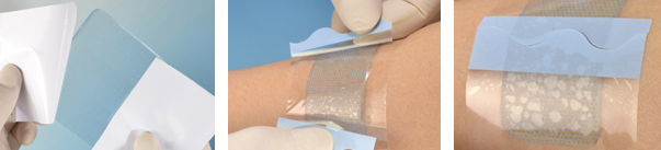
Inspired by the octopus's suckers, researchers have developed a personalized, suction-based adhesive patch with selective adhesion and stretchability capabilities. These patches are constructed using silicone rubber membranes, featuring microscopic sucker structures that can adhere to normal skin while their biocompatible Gelatin Methacryloyl (GelMA) hydrogel interfaces with injured areas. The sucker microstructures on the silicone rubber film enable these patches to exhibit strong adhesion to both dry and wet surfaces.
By incorporating Vascular Endothelial Growth Factor (VEGF) into the GelMA hydrogel, the healing process can be further accelerated, and the shape of the GelMA hydrogel can be customized to fit the geometry of the wound area. Integrating both adhesive and anti-adhesive properties into a single patch membrane allows for precise coverage of individual wound regions and has been shown to effectively promote wound healing in rat skin defect models.
The development of medical adhesives utilizing the octopus's suction mechanism avoids the adverse effects associated with chemical adhesion. However, the reported octopus-inspired adhesion system requires an additional negative pressure generation system to reduce the pressure within the suckers. Moreover, the current imitation of the octopus's sucker structure is limited to adhering to smooth surfaces, whereas most human tissues and wounds possess millimeter/micrometer-scale protrusions, curvatures, and angles.
4.Slug
The slug, commonly known as a "snail without a shell," is a hermaphroditic mollusc. When threatened, the slug secretes a copious amount of mucus on its dorsal surface, adhering itself to rocks and soil, making it difficult for predators to carry it away, thus achieving self-defense. The mucus of the slug forms a highly resilient double network crosslink through the interaction of long, intertwined, negatively charged heparan sulfate-like polysaccharides and positively charged proteins.
Inspired by the slug's mucus, researchers have developed a double-layered adhesive consisting of a surface layer and a dissipative matrix. The surface of this adhesive adheres firmly to tissues through electrostatic interactions, covalent bonds, and physical penetration, demonstrating potential as a high-performance skin adhesive in vitro studies for skin adhesion applications.
5.Gecko
The gecko, a type of lizard, is renowned for its ability to move freely on walls, thanks to the approximately 500,000 hair-like fibrils, known as setae, densely packed in an array on each foot pad. Each seta is further subdivided into hundreds of tiny, spatula-shaped nanostructures with tips measuring approximately 200-500 nanometers in length. These tips interact with surfaces of various shapes through van der Waals forces and capillary action, enabling the gecko to generate a remarkable adhesion force through this hierarchical structure. Gecko adhesive possesses anisotropic adhesion, a high adhesion coefficient, low peeling force, material-independent adhesion, self-cleaning capabilities, anti-self-adhesion, and a non-sticky default state.
Inspired by the gecko's adhesion mechanism, dry medical patches have been developed to replace existing pressure-sensitive adhesives and wet acrylic medical patches, offering self-cleaning and reusable adhesion properties. Furthermore, gecko-inspired medical adhesives for wet tissue environments have also been created, including one based on polyglyceryl sebacate acrylate (PGSA), which exhibits elasticity and biodegradability. This tissue adhesive is optimized for adhesion by altering the dimensions of the nanoscale pillars, including the ratio of tip diameter to pitch and tip diameter to base diameter.
Most research on gecko-like adhesives focuses on rod- and wire-shaped features with high aspect ratios (AR). However, solely modifying surface structures by varying the aspect ratio to mimic gecko adhesion is challenging. An alternative approach involves coating the material surface with an embossed film, which can alter the surface's adhesion properties.
6.Giant Salamander
The giant salamander, commonly known as the baby fish, is a precious amphibian unique to China. Its skin mucus glands secrete a white mucus when subjected to external stimuli, which aids the giant salamander in evading danger and promoting wound healing. This mucus contains 155 types of peptides/proteins, with amino acids containing phenyl rings accounting for over 11% (tyrosine at 5.59%, phenylalanine at 5.08%, and tryptophan at 0.94%). The high activity of phenolic hydroxyl groups participating in hydrogen bonding and π-π conjugation contributes to its tissue adhesion properties. Additionally, physical interactions such as hydrophobicity/hydrophilicity, hydrogen bonding, and ionic complexation also contribute to its ability to adhere in wet environments.
When used as a tissue adhesive, the freeze-dried powder of the giant salamander's mucus (SSAD, Andrias davidianus), after cleaning and separation, has been shown to promote faster and better healing of skin incisions on the backs of rats compared to commercially available fibrin glue and cyanoacrylate glue. Furthermore, it can serve as an effective dressing to promote healing of skin defects in diabetic rats.
References
[1] Che Chaoyue, Yuan Kunshan, Zhang Shuxin, Zhang Haijun. Research Progress of Medical Biomimetic Adhesives. China Adhesives, 2021, 30(10): 62-66. DOI: 10.13416/j.ca.2021.10.013.
[2] Chen Yuqin, Wang Xiaoping, Zhang Ximu. Research Progress of Biomimetic Natural Product-Based Medical Adhesives for Skin Wound Healing. Chinese Journal of Medical Aesthetics and Cosmetology, 2023, 13(01): 49-52. DOI: 10.19593/j.issn.2095-0721.2023.01.012.
[3] Wu Yun, Li Jian. Research Progress of Aquatic Organism-Inspired Medical Adhesives. Journal of Biomedical Engineering, 2019, 36(02): 325-333.
About the Author
Xiaonisha, a food technology professional holding a Master's degree in Food Science, is currently employed at a prominent domestic pharmaceutical research and development company. Her primary focus lies in the development and research of nutritional foods, where she contributes her expertise and passion to create innovative products.


 ALL
ALL Pharma?in?China
 Pharma?Experts
 Market News
 Products Guide
 Brand Story


 Pharma Sources Insight July 2025


