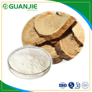
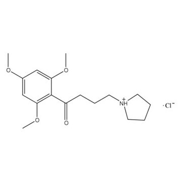
Oxymatrine is a natural alkaloid primarily found in Sophora flavescens Ait., Sophora tonkinensis Gagnep., Sophora alopecuroides L., and Sophora viciifolia Hance, all belonging to the Fabaceae family. A considerable number of studies have found that oxymatrine can counteract the damage to vascular endothelial cells caused by hypoxia, high glucose, and bacterial toxins, inhibit the endothelial-mesenchymal transition, and also combat hypoxia, TGF-β (transforming growth factor-β), and hyperlipidemia-induced proliferation of vascular smooth muscle cells as well as the damage to vascular smooth muscle cells caused by β-glycerophosphate. This results in vascular protection and improved vascular function. Oxymatrine can dilate blood vessels and counteract the vasoconstrictive effects of high glucose, high fat, and phenylephrine on blood vessels. It can also produce a hypotensive effect by blocking nerve endings and promoting the synthesis and release of nitric oxide. Additionally, it can regulate blood pressure by improving cardiac function. Oxymatrine can inhibit pulmonary hypertension and pulmonary vascular remodeling induced by hypoxia or monocrotaline in rats, as well as atherosclerosis caused by a high-fat diet.
Protecting Vascular Endothelial Cells
Vascular endothelial cells possess various crucial physiological functions, such as regulating vascular tone, maintaining the balance between coagulation and fibrinolysis systems, modulating the aggregation of inflammatory cells, and inhibiting platelet aggregation. Endothelial cell injury and dysfunction are the initiating steps in the formation of atherosclerotic lesions, and they are closely related to myocardial ischemia in coronary heart disease. Therefore, protecting vascular endothelial cells is an important strategy for preventing and treating cardiovascular diseases. Oxymatrine has the effect of protecting various vascular endothelial cells.
1.Protecting Umbilical Vein Endothelial Cells
In experiments studying the anti-tumor angiogenesis effects of oxymatrine in vitro, human umbilical vein endothelial cells were used as a control. The results showed that the half-maximal inhibitory concentration (IC50) of oxymatrine in inhibiting the proliferation of human umbilical vein endothelial cells was 1555 mg/L. Oxymatrine could cause deep nuclear staining, reduced cell volume, irregular cell shape, decreased extracellular villi, and most cells were in an early apoptotic state. It significantly inhibited the activity of succinate dehydrogenase in aerobic oxidation. Oxymatrine could increase the survival rate of human umbilical vein endothelial cells impaired by hypoxia-reoxygenation in a concentration-dependent manner, reduce the content of lactate dehydrogenase in the culture medium, upregulate the Bcl-2/Bax ratio, and downregulate the expression of caspase-3 protein. Additionally, some studies have reported that oxymatrine concentrations of 1 and 3 μmol/L did not affect the proliferation of human umbilical vein endothelial cells, while concentrations of 10 and 30 μmol/L inhibited their proliferation. At 3 μmol/L, oxymatrine could counteract the damage and reduced activity of human umbilical vein endothelial cells caused by high glucose. It could also counteract the upregulation of A2B adenosine receptor and independent chemokine CCL5 receptor expression, as well as the phosphorylation of p38 and extracellular signal-regulated kinase 1/2 (ERK1/2) caused by high glucose. This indicates that the mechanism of oxymatrine in counteracting high glucose-induced endothelial cell damage is through downregulating the expression of A2B receptor.
2.Protecting Arterial and Microvascular Endothelial Cells
Oxymatrine, at low concentrations, has a promoting effect on endothelial cell proliferation, which is related to inhibiting the secretion of vascular endothelial growth factor by tumor cells and downregulating the expression of growth factor receptors related to blood vessels. According to studies, continuous intraperitoneal injection of oxymatrine at 120 mg/kg for 10 days could reduce the protein expression of nuclear factor-κB (NF-κB), ERK1/2, p38MAPK, and JNK in the basilar artery endothelial cells of rats with subarachnoid hemorrhage, as well as the gene expression of interleukin (LT)-1β, IL-6, and tumor necrosis factor-α (TNF-α). The related mechanism is related to downregulating the expression of A2B receptor, blocking the MAPK/ERK/JNK signaling pathway, and inhibiting inflammatory responses. Additionally, oxymatrine can inhibit endothelial-mesenchymal transition and protect vascular endothelial cells by downregulating the expression of hypoxia-inducible factor-1α, thus inhibiting vascular remodeling.
Inhibiting Vascular Smooth Muscle Cell Proliferation
Oxymatrine can concentration-dependently inhibit the incorporation of 3H-thymidine into normal aortic smooth muscle cells and those obtained from a rabbit model of atherosclerosis induced by high-fat diet, thereby inhibiting DNA synthesis in smooth muscle cells. It can also inhibit the proliferation of aortic smooth muscle cells and the gene expression of c-fos and c-myc in the model rabbits, causing cell cycle arrest in G1 and G2+M phases, with a decrease in S-phase cells. Oxymatrine significantly inhibits the increased proliferation of primary rat pulmonary artery smooth muscle cells induced by transforming growth factor-β (TGF-β) or hypoxic culture. It counteracts the upregulation of reactive oxygen species and H2O2 generation in pulmonary artery smooth muscle cells induced by hypoxia, as well as the downregulation of reduced glutathione levels. The mechanism involves upregulating the protein expression of nuclear factor erythroid 2-related factor 2 (Nrf2), thereby upregulating the expression of antioxidant proteins superoxide dismutase-1 (SOD-1) and heme oxygenase-1 (HO-1), exerting anti-hypoxia-induced oxidative stress injury. Oxymatrine can concentration-dependently counteract the calcification induced by β-glycerophosphate, reducing calcium content and malondialdehyde content in arterial smooth muscle cells, and decreasing alkaline phosphatase activity. Its mechanism is related to blocking the TGF-β/Smads/core binding factor-α1 (Cbfα1) signaling pathway and upregulating the expression of osteoprotegerin while downregulating the expression of NF-κB receptor activator ligand (the pseudoreceptor of osteoprotegerin).
Protecting Blood Vessels and Regulating Their Contraction, Dilation, and Blood Pressure
1. Protecting Blood Vessels and Inhibiting Angiogenesis
Studies have reported that continuous intraperitoneal injection of oxymatrine at 100 mg/kg for 27 days in a rat model of pulmonary fibrosis induced by bleomycin significantly reduced alveolar inflammation and fibrous collagen tissue proliferation in the lung tissue of the model rats. During the early stages of pulmonary fibrosis, it significantly counteracted the increase in lung microvascular density caused by bleomycin. This mechanism is related to inhibiting neovascularization in the early stages of pulmonary fibrosis, correcting the imbalance between Th1 and Th2 cells, and inhibiting collagen synthesis.
2. Improving Vascular Contraction and Dilation Functions to Regulate Blood Pressure
Oxymatrine possesses anti-inflammatory properties that protect vascular endothelial cells and alleviate cerebral vasospasm in rats with subarachnoid hemorrhage. Intravenous injection of oxymatrine at 4 mg/kg in mice significantly increased the diameter, flow velocity, and flow volume of cerebral microvasculars, indicating that oxymatrine not only dilates microvessels but also enhances the amplitude and frequency of the autoregulatory movement of microarteries. This activation of the inhibited autoregulatory movement of microarteries regulates microvascular function, prevents blood cell aggregation, thereby redistributing local blood flow, improving blood perfusion, and oxygen exchange. Continuous administration of oxymatrine at 100 mg/kg for 4 weeks by gavage in type 2 diabetic rats established by feeding a high-sugar, high-fat diet combined with low-dose streptozotocin injection significantly increased the percentage of thoracic aortic dilation in the model rats, reduced the elevated gene expression of A2B receptors in the aorta, and significantly lowered the mean blood pressure, systolic blood pressure, and diastolic blood pressure in the model rats. This dosage did not affect fasting or postprandial blood glucose levels in the model rats.
When administered to rats with heart failure induced by isoproterenol, oxymatrine can improve heart failure while upregulating the protein expression of endothelial nitric oxide synthase (eNOS) and NO levels in the myocardium. Oral administration of oxymatrine can also upregulate the expression of eNOS and phosphorylated eNOS in the lung tissue of rats with pulmonary hypertension, promote the catabolism of asymmetric dimethylarginine (ADMA), which is a competitive inhibitor of eNOS, restore and accelerate NO synthesis. This suggests that oxymatrine can dilate blood vessels by promoting NO synthesis.
Anti-Pulmonary Hypertension and Anti-Atherosclerosis
1. Anti-Pulmonary Hypertension Effect
Using monocrotaline to create a rat model of pulmonary hypertension, continuous administration of oxymatrine by gavage for 28 days significantly reduced the lung index and right ventricular hypertrophy in the model rats, improved right ventricular contractile function (reduced right ventricular systolic pressure and maximum rate of change in right ventricular internal pressure), without affecting heart rate, and reversed right ventricular hypertrophy. It also reduced serum cardiac troponin-I levels in rats with pulmonary hypertension, improved the structure of small pulmonary arteries, reduced wall thickness, increased lumen area, reduced inflammatory cell infiltration, improved pulmonary arteriolar remodeling, decreased pulmonary vascular resistance, lowered overall pulmonary vascular resistance, reduced pulmonary artery pressure, relieved right ventricular afterload, and reversed right ventricular failure. Studies on the mechanism of action found that oxymatrine reduced serum ADMA levels in model rats, upregulated the protein expression of protein kinase B (Akt) and phosphorylated Akt in lung tissue to levels similar to those in normal rats. It also upregulated the expression of eNOS, phosphorylated eNOS, and dimethylarginine dimethylaminohydrolase-2 (DDAH2) in lung tissue. Since it did not affect the expression of protein arginine methyltransferase-1, it is believed that oxymatrine promotes the catabolism of eNOS's competitive inhibitor ADMA, restores and accelerates NO synthesis, and produces an anti-pulmonary hypertension effect by promoting the Akt/eNOS signaling pathway and upregulating the expression of DDAH2 in lung tissue.
When rats were placed in a hypoxic chamber to create a model of hypoxic pulmonary hypertension, intraperitoneal injection of oxymatrine before daily hypoxic exposure significantly reduced right ventricular systolic pressure, the ratio of right ventricular weight to left ventricular plus interventricular septum weight, and right ventricular hypertrophy. It also significantly alleviated pathological changes such as reduced thickness of small pulmonary vessel walls and inflammatory cell infiltration, thus mitigating small pulmonary vessel remodeling. Oxymatrine reduced the contractile response of pulmonary artery rings in hypoxic pulmonary hypertension rats to phenylephrine. It counteracted the overexpression of inflammatory-related factors such as monocyte chemoattractant protein-1, IL-6, stromal cell-derived factor-1, vascular cell adhesion molecule-1, and intercellular adhesion molecule-1, as well as the overexpression of NF-κB protein and genes related to cell growth and differentiation, including TGF-β, vascular endothelial growth factor, and endothelin-1, in the lung tissue of the model rats. Oxymatrine also counteracted the protein expression of monocyte/macrophage surface antigen ED-1 and T lymphocyte surface antigen CD3 in the lung tissue of the model rats, inhibiting the accumulation of monocytes/macrophages and T lymphocytes around the pulmonary blood vessels caused by hypoxia, resulting in normalization of alveolar tissue, significant reduction of inflammatory cell infiltration, and attenuation of small vessel wall hypertrophy. Mechanism studies suggest that oxymatrine achieves these effects by activating the Nrf2/HO-1 and Nrf2/SOD-1 pathways, initiating the inhibition of endothelial-mesenchymal transition in endothelial cells, thereby alleviating hypoxia-induced pulmonary vascular remodeling.
2. Anti-Atherosclerosis Effect
Oxymatrine has the ability to inhibit the formation of arterial lipid plaques. It can lower the total cholesterol level in the serum of model mice, increase the high-density lipoprotein cholesterol level, without affecting the levels of triglycerides and low-density lipoprotein cholesterol. It also downregulates the gene and protein expression of Yes-associated protein (YAP) in the aorta, and upregulates the gene and protein expression of retinoic acid receptor-alpha (RARα), ATP-binding cassette transporter (ABC) A1, and ABCG1. Oxymatrine can concentration-dependently reduce the levels of total cholesterol, free cholesterol, and cholesterol esters in foam cells, promoting the efflux of cholesterol from the cells to apolipoprotein A-1 (ApoA-1) and high-density lipoprotein. Oxymatrine also downregulates the gene and protein expression of YAP, and upregulates the gene and protein expression of RARα and RARγ within the retinoid acid receptor family. In summary, oxymatrine inhibits the development of atherosclerosis by inhibiting the gene and protein expression of YAP in macrophages (foam cells), upregulating the gene and protein expression of RARα, ABCA1, and ABCG1, promoting cholesterol efflux from cells, reducing intracellular cholesterol concentration.
References
[1] Wang Cheng, Zhang Mingfa, Shen Yaqin. Research Progress on Vascular Pharmacological Effects of Oxymatrine [J]. Drug Evaluation Research, 2021, 44(07): 1555-1561.
About the Author
Xiaomichong, a researcher in pharmaceutical quality, has been dedicated to pharmaceutical quality research and verification of drug analysis methods for a long time. Currently, she works in a large domestic pharmaceutical research and development company, engaged in drug inspection analysis and verification of analytical methods.


 ALL
ALL Pharma?in?China
 Pharma?Experts
 Market News
 Products Guide
 Brand Story


 Pharma Sources Insight July 2025


